| Issue |
Int. J. Lim.
Volume 59, 2023
|
|
|---|---|---|
| Article Number | 5 | |
| Number of page(s) | 8 | |
| DOI | https://doi.org/10.1051/limn/2023006 | |
| Published online | 28 June 2023 | |
Research article
Effects of ABS microplastics on microalgae Chlorella vulgaris and Raphidocelis subcapitata
1
Institute of Science and Technology, São Paulo State University, UNESP, 3 de Março Avenue 511, Alto da Boa Vista, 18087-180 Sorocaba, Brazil
2
Department of Ecology, Institute of Biosciences, University of São Paulo, USP, Matão Street 321, 05508-090 São Paulo, Brazil
3
Department of Microbiology and Ecology, Cavanilles Institute of Biodiversity and Evolutionary Biology, University of Valencia, Catedràtic José Beltrán Martínez, 2, 46980 Paterna, Spain
4
Department of Fundamental Chemistry, Institute of Chemistry, University of São Paulo, USP, Prof. Lineu Prestes Avenue 748, 05508-000 São Paulo, Brazil
* Corresponding author: This email address is being protected from spambots. You need JavaScript enabled to view it.
Received:
3
November
2022
Accepted:
8
June
2023
Abstract
In recent years, there has been a growing interest in the impacts caused by the presence of microplastics (MP) in aquatic environments. The impacts of microalgae exposure to microplastics are still insufficiently investigated and further studies are needed to understand the possible outcomes. In addition, much of the literature has focused on the study of concentrations above those found naturally in the environment and in less toxic polymer matrices. Acrylonitrile-butadiene-styrene (ABS) plastics have a composition rich in additives and, so far, have been studied superficially. In the present study, two of the most commonly used green microalgae species in toxicity assays, Chlorella vulgaris and Raphidocelis subcapitata, were exposed to different concentrations of primary ABS-MP for a period of 6 days. Here, we observed physiological changes in cell growth and chlorophyll a content induced by the concentration and time of exposure to ABS-MP. The lowest concentration did not prove to be potentially toxic to cells, while the highest concentration was the most toxic. Primary consumers, such as microalgae, are essential for the proper functioning of entire ecosystems. Changes in these communities can lead to permanent damage to the communities of organisms at higher levels, so it is essential that their study be done carefully in the face of threats such as MP.
Key words: Biological effects / ecosystem effects / plastic pollution / pollutants / toxicity
© EDP Sciences, 2023
1 Introduction
The perception of the gravity of microplastics (MP) pollution is recent. Initially focused on marine environments, the studies have also investigated freshwater environments, such as lakes, rivers, and reservoirs (Buteler et al., 2023; Cincinelli et al., 2017; Luo et al., 2019). Freshwater bodies close to human-occupied areas are more susceptible to MP pollution due to the release of domestic and industrial wastewater and inadequate disposal of large plastic materials (Xu et al., 2020).
MP can remain in the water column and bottom sediment in the aquatic environment. Disponible in these compartments, plastic particles can interact and be ingested by organisms of different trophic levels (Ain Bhutto and You, 2022; Garcia et al., 2021; Krause et al., 2021), such as microalgae (Chen et al., 2020; Déniel et al., 2020; Hazeem et al., 2020), crustaceans (Canniff and Hoang, 2018; Portugal et al., 2021), insects (Carrasco-Navarro et al., 2021; Ribeiro-Brasil et al., 2022), mollusks (Esterhuizen et al., 2022; Hoellein et al., 2021; Wang et al., 2022), and fish (Abarghouei et al., 2021; Batel et al., 2020; Zhang et al., 2021). However, algae represent a group superficially evaluated by studies that investigate the interaction of MPs with aquatic species.
Microalgae are organisms with extremely varied characteristics that can be found in almost all aquatic environments (Li et al., 2022; Mutanda et al., 2020). As primary producers, these organisms play a key ecological role in maintaining food chains. For this reason, these organisms have been widely used in toxicological tests. However, due to the great variability of algae characteristics, different responses can be observed according to species and pollutant (Song et al., 2020).
Many studies have reported the reduction in algae growth after exposure to different types of microplastics (MP), such as polystyrene (PS) (Casado et al., 2013), polyvinyl chloride (PVC) (Fu et al., 2019), polyacrylonitrile (PAN) (Lin et al., 2020), polypropylene (PP) (Wu et al., 2019), polyamide (PA) and polyethylene (PE) (Yang et al., 2020). Other outcomes were also studied, such as oxidative stress, enzyme activity, photosynthetic activity and pigment production (Rani-Borges et al., 2021). However, studies at environmentally relevant concentrations are still not common. As well as studies that consider more complex scenarios, such as the interaction with two or more pollutants, and more specific cellular impacts, such as the cellular internalization of particles, which according to Chen et al. (2020) can trigger negative effects, such as altering fertility and algal growth.
In the literature, significant effects have been observed in the different species of microalgae studied, although the results are not consistent in all studies. Many authors reported that no negative effects were found after the exposure period or even that population parameters benefited from the presence of MP in the medium, such as cell growth (Canniff and Hoang, 2018; Song et al., 2020; Yokota et al., 2017). Furthermore, according to the study by Song et al. (2020), although there were moderate changes in the chlorophyll a content, the differences were not statistically different from the control group. Overall, these mixed results suggest that the effects of microplastic exposure on microalgae are not yet well-established, and further research is needed to understand the underlying patterns of these interactions.
The study of less produced polymers, but with high toxic potential, has also been neglected. The use of copolymers by the industry has been increasingly frequent, since it is a way to obtain a final product with different characteristics and properties through the joining of two structurally distinct homopolymers (Tager, 1972). Acrylonitrile-butadiene-styrene (ABS) copolymer is a polymer widely used in building materials, electronics, automotive and transportation industries (Li et al., 2017; Mao et al., 2016; Scaffaro et al., 2012). According to the risk assessment of polymers, ABS is highly toxic, mainly due to the presence of endocrine disruptors in the polymer matrix (Lithner et al., 2011), which constitutes a major threat to aquatic biota. Furthermore, ABS microplastics (ABS-MP) have been detected in freshwater samples from different locations, indicating its wide environmental distribution. It has been found in drinking water treatment plants (Siegel et al 2021), groundwater (Kim et al 2023), surface water samples and sediment samples (Drabinski et al 2023; Li et al 2021), and internalized by species of insect larvae (Akindele et al 2020; Ehlers et al 2019).
Considering previous studies that show inconsistent results regarding the toxicity of MP to microalgae cells, the general objective of this study was to investigate the biological response of algae to a copolymer harmful to many species. The study of this interaction becomes a relevant source of ecological data, since algae play a fundamental role in the metabolism of the aquatic ecosystem. The species studied were Chlorella vulgaris and Raphidocelis subcapitata. Cultures were exposed for 6 days (chronic exposure) to three concentrations of ABS-MP and analyzed for growth inhibition and chlorophyll a content.
2 Material and methods
2.1 Microplastic characterization
The MP used in the experiment was composed of primary ABS, with a size <100 μm, irregular shape and black color. The ABS sample was donated by a local industry. ABS was selected for this study due to its widespread use across various industrial sectors (Li et al., 2017; Mao et al., 2016; Scaffaro et al., 2012), and it is a poorly studied polymer, despite its high toxicity, being considered one of the five most toxic polymers in the world (Lithner et al., 2011). Therefore, it is important to carry out studies that can assess the responses of primary producers to this type of MP.
The polymeric composition of ABS-MP was confirmed using a micro-Fourier transform infrared spectroscopy (micro-FTIR). The FTIR spectra were measured in a Bruker spectrometer, Alpha model, in the region of 400–4000 cm−1, with standard KBr beamsplitter and high sensitivity DLATGS detector. The spectra were recorded with the ATR (Attenuated Total Reflection) module: ATR Platinum, equipped with a diamond crystal as a reflective element. The spectra were obtained with 128 accumulations and with a resolution of 2 cm−1 (Fig. 1A).
The MP was acquired in the form of powder and subsequently subjected to cryolamination. For a better visualization of the particles and to confirm the irregular shape of the particles, ABS-MP was stained with Nile Red and observed under an inverted fluorescence microscope (Leica DMi8) (Fig. 1B). The confirm size range of the particles and to determine the MP concentrations used in the toxicity tests, the Zeiss Discovery V12 stereomicroscope was used to count the particles present in 30 μL of the MP solution. The samples were previously vacuum filtered and the count was performed in four repetitions and the mean and standard deviation were calculated. For every 30 μL of the MP solution we found 196 ± 11 items. The selection of concentrations was based on studies conducted with environmental samples of freshwater. As demonstrated in the extensive review by Koelmans et al. (2019), reported MP concentrations vary significantly between different locations, spanning ten orders of magnitude (ranging from 1×10⁻² to 108/m3). Taking this into account, we used a minimum concentration of 384 items L−1. For the medium and higher concentrations, we employed 3840 and 38400 items L−1, respectively, which are 10 and 100 times higher, aiming to induce more pronounced physiological responses.
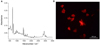 |
Fig. 1 Fluorescence image of acrylonitrile-butadiene-styrene microplastic (ABS-MP) confirming the irregular shape of the particles. |
2.2 Microalgae species
The species studied were Chlorella vulgaris and Raphidocelis subcapitata, both green microalgae commonly used in toxicity assays. The common use of Chlorella is mainly due to the fact that this genus is considered ideal for commercial and biotechnological purposes, due to its easy reproduction in the presence of sunlight, carbon dioxide, water and small amounts of nutrients, facilitating its cultivation (Masojídek and Torzillo, 2008). Furthermore, the ability to accumulate high concentrations of carotenoids makes Chlorella sp. even more interesting for its use in biotechnology. Regarding its shape, Chlorella has a spherical shape, however, as the cells grow, its structural shape changes.
R. subcapitata belongs to the unicellular freshwater Chlorophyceae class, known to be more susceptible to chemical compounds than other species, so it is recommended for ecotoxicological assays (OECD, 2011). Its cultivation does not require many requirements, its growth rate is quite high and its genome has already been sequenced, such characteristics make this species ideal to be applied in several studies (Almeida et al., 2019).
2.3 Experimental design and analysis
The green microalgae C. vulgaris and R. subcapitata were cultivated in NPK culture medium (10-5-10 g L−1) in an incubator with continuous lighting (1200–1900 lux), with a photoperiod of 12:12 hours (light:dark) and temperature of 21 ± 2°C. The experiment was started when the cultures were in the exponential growth phase. The experiments were carried out in glass Erlenmeyer flasks containing 500 mL of culture of both species. The test was performed over a period of 6 days. Each treatment and negative control were performed in triplicate (n = 3, N = 12). Cultivation conditions were maintained during the experimental period, with the exception of aeration.
ABS-MP were added directly to the culture medium. The concentrations tested were Concentration 1: 384 items L−1; Concentration 2: 3840 items L−1 and Concentration 3: 38,400 items L−1.
The toxicity of MP exposure was measured by analyzing cell growth and chlorophyll a concentration (Strickland and Parsons, 1972). Analyzes of growth and pigment content were performed on the initial (day 0) and final (day 6) days of exposure. The growth of the algal culture was calculated by counting cells in a Neubauer chamber under light microscopy with the aid of a manual counter. For pigment analysis, 50 mL of the culture was vacuum filtered through glass fiber filters (Millipore 47 mm with a pore size of 0.7 μm). The filters were placed in 15 mL tubes and frozen at −20 °C protected from light until processing. 24 hours before the procedure, 5 mL of DMSO + 90% acetone solution was added to each tube and stored at 4 °C. After this period, the samples were centrifuged (3500 rpm, 4 °C for 10 min) and the supernatant was transferred to a 5 mL cuvette for reading in a spectrophotometer (Femto, Cirrus 80) at 433, 480, 510, 647, 667 and 750 wave length.
The results of algae growth and chlorophyll a content were displayed as a percentage (%) calculated from the results obtained from the results of control group at each sampling time (days 0 and 6).
2.4 Statistical analysis
To determine whether there were significant differences between treatments (different concentrations of MP) and the control group, and between treatments according to exposure time, Dunnett's and Fisher's exact statistical tests were used, respectively, in addition to descriptive analyzes (mean and relative standard deviation). All analyzes were performed in triplicate and statistical significance was accepted at p < 0.05 level. The software used was Minitab v.14.
3 Results and discussion
3.1 Growth inhibition
After 6 days of exposure to ABS-MP, samples of C. vulgaris and R. subcapitata were analyzed to assess the effects of this pollutant on cell growth.
The growth of C. vulgaris (Fig. 2A) remained stable at all concentrations tested, when compared to the control group (Dunnett's test, p < 0.05). At Concentration 2 (3840 items L−1), growth was inhibited at the end of the 6-day exposure period (Fisher test, p < 0.05). In treatments with R subcapitata (Fig. 2B), cell growth was observed in relation to the control group only at Concentration 2 (3840 items L−1) after 6 days of exposure (Dunnett test, p < 0.05), which also differed in relation to the first timepoint (Day 0) (Fisher test, p < 0.05).
The stability in cell number observed for both species in the treatment under Concentration 1, shows that ABS-MP in this amount of particles (384 items L−1), did not show toxicity at the population level. Lagarde et al. (2016) reported in their 78-day study that the culture of Chlamydomonas reinhardtii did not undergo any change in its growth pattern before 63 days of exposure to polyethylene and to polypropylene MP. Despite this, the concentration used in this study was much higher than that used in the present work, therefore, it is not possible to infer whether in a long exposure the results would be different. Casado et al. (2013) used R. subcapitata with realistic concentrations and observed a reduction in growth, however, the physical characteristics of the MP used were quite different from the ABS-MP used here, being ∼11 μm. Smaller particles are known to generally have greater toxicity. All studies found in the literature with C. vulgaris used concentrations considered high (>10 mg L−1), making comparison with our results impossible (Fu et al., 2019; Hazeem et al., 2020; Sjollema et al., 2016; Song et al., 2020).
In treatment with C. vulgaris under Concentration 2, cells were significantly inhibited when compared with day 0 (p < 0.05). This result can be attributed to a combination of factors that induce different responses in cells, such as the shading effect, increased turbidity of the medium, cell internalization process and adhesion of MP to the cell wall, which inhibit cell growth (Lagarde et al., 2016). After these processes, cells can undergo a period of adaptation and self-regulation, returning to the state of exponential growth (Rani-Borges et al., 2021). To observe this event, studies with longer exposure times should be performed. Furthermore, the toxic potential of ABS comes mainly from the release of additives present in its polymer matrix (Lithner et al., 2011). Therefore, it is to be expected that the exposure time is a determining factor for the increase in the toxicity of this material.
On the other hand, R. subcapitata showed growth at Concentration 2, both compared with Day 0 and control treatment (p < 0.05). This phenomenon was also observed by Canniff and Hoang (2018) with the same species exposed to polyethylene (PE) MP with a size range within the one used here (63–75 μm). It is suggested that this event occurs due to the surface of MP that can serve as a substrate for the adhesion and growth of microorganisms, which favors the increase of algae concentration (Canniff and Hoang, 2018; Song et al., 2020; Yokota et al., 2017).
It is noteworthy that the size of MP particles can be an important factor of toxicity. The microalgae Skeletonema costatum, when exposed to PVC particles of 1 µm, in concentrations between 20 and 200 mg L−1, had its cell growth significantly inhibited. The same does not occur when exposed to 1 mm PVC particles (Zhang et al., 2017). The growth of the microalgae Dunaliella tertiolecta also seems to be affected by particle size. MP-PE caused a more significant inhibitory effect with smaller particle sizes (Sjollema et al., 2016). The fact that smaller particles can be more toxic is very worrying, especially if we consider that, as the plastic material remains in the environment, smaller and smaller particles are formed and, consequently, can increase the polluting potential of these contaminants.
Overall, the rate of algal growth inhibition is one of the most used parameters in laboratory ecotoxicological studies using algal cultures. These experiments allow us to suggest the risks of a xenobiotic on an algae population and, consequently, to estimate the impact of this contaminant on the entire ecosystem, since algae constitute a group of extreme importance for the maintenance of balance in aquatic environments.
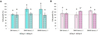 |
Fig. 2 Cell growth (%), normalized to the negative control group (100%, represented by the red dashed line), and relative standard deviation of days 0 (initial) and 6 (final) of exposure to acrylonitrile-butadiene-styrene microplastics (ABS-MP) at three concentrations: 384, 3840 and 38,400 items L−1. (A) Chlorella vulgaris (in green). (B) Raphidocelis subcapitata (in pink) Equal letters indicate no significant difference and different letters indicate a significant difference between the groups at different exposure times (days 0 and 6) (Fisher's exact test). The asterisk symbol (*) represents a statistically significant difference between the treatments and the control group (Dunnet's test). p value considered significant p < 0.05. |
3.2 Chlorophyll a
Chlorophyll a is a photosynthetic pigment located in chloroplast membranes that absorbs light energy to be used in photosynthesis, a fundamental process for the biological activities of microalgae. Chlorophyll a content was measured at the beginning of the exposure period (day 0) and at the end (day 6) in order to evaluate the effects of ABS-MP on the production of this pigment.
The species C. vulgaris did not show significant differences (Fisher test, p < 0.05) in any of the concentrations tested between days 0 and 6 of exposure. However, the culture exposed to Concentration 3 was statistically different from the control group (Dunnett's test, p < 0.05) at both evaluated times (Fig. 3A). On the other hand, R. subcapitata showed an increase and decrease in chlorophyll a content under Concentrations 1 and 3, respectively (Fisher test, p < 0.05), after 6 days of exposure (Fig. 3B). When compared to the control group, R. subcapitata showed a significant reduction at the beginning of the experiment (day 0) at all concentrations tested, and at day 6 at the highest concentration (38,400 items L−1) (Dunnett test, p < 0 .05).
In the experiments with C. vulgaris there was no significant difference over the 6-day exposure period (Fisher test, p < 0.05). With the exception of Concentration 3, which had production inhibited, the groups exposed to Concentrations 1 and 2 did not differ from the control (Dunnett test,p < 0.05). Song et al. (2020) reported a similar result in a study with Chlorella sp., after tests under different experimental conditions (concentration of 200 mg L−1 for 96 h) and MP consisting of other polymers (PE, PET, PP and PVC; with a size of 74 μm). On the contrary, Tunali et al. (2020) demonstrated a reduction in chlorophyll production by C. vulgaris after exposure to polystyrene MP (0.5 µm) and that the reduction was accentuated as the concentration increased. Thus, with these results it is possible to suggest that the physiological responses of pigment production will be intrinsic to the physical and chemical characteristics of the particles to which the alga will be exposed as well as the species itself and the particle size. All these parameters may be a fundamental factor for the understanding of the toxic effects of MP on algae.
Concerning R. subcapitata, curiously, under Concentration 1 shows a significant increase in the production of chlorophyll a (Fig. 3B), even though there was no cell growth in this same treatment (Fig. 2B). At the same time, under Concentration 2, no increase in chlorophyll a production was observed, even though there was an increase in cell growth (Fig. 2B). This suggests that the lower concentration may not have caused damage at the physiological level, while under 3840 items L−1 the photosynthetic apparatus suffered some damage, even with the increase in population. These conclusions should be taken into account with caution, as there was a reduction in chlorophyll a content at all concentrations tested on day 0 compared to the control group (equivalent to 100% chlorophyll content).
The greatest vulnerability of R. subcapitata to the presence of ABS-MP occurred in the exposure of ABS-MP to 38,400 items L−1, with a reduction of pigment production of ∼12% in 6 days of experiment. Although lower concentrations than those normally found in the literature were used, the results demonstrate the same pattern observed in other studies, where the rate of chlorophyll inhibition was dose-dependent (Li et al., 2020; Tunali et al., 2020; Wang et al., 2020; Wu et al., 2019). However, in the present study, this effect was observed only for R. subcapitata.
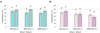 |
Fig. 3 Chlorophyll a content (%), normalized with the negative control group (100%, represented by the red dashed line), and relative standard deviation of days 0 and 6 of exposure to acrylonitrile-butadiene-styrene microplastics (ABS-MP) at three concentrations: 384, 3840 and 38400 items.L−1. (A) Chlorella vulgaris (in green) (B) Raphidocelis subcapitata (in pink). Equal letters indicate no significant difference and different letters indicate a significant difference between the groups at different exposure times (days 0 and 6) (Fisher's exact test). The asterisk symbol (*) represents a statistically significant difference between the treatments and the control group (Dunnet's test). p value considered significant p < 0.05. |
3.3 Environmental implications of the interaction between MP and microalgae
The effects observed through experiments with C. vulgaris and R. subcapitata could potentially have ecosystem implications. However, it is difficult to predict how these effects might manifest under real conditions in freshwater environments due to the complexity of these ecosystems. Microalgae represent a key group for maintaining ecological balance in aquatic environments (Wu et al., 2017). The changes caused by increasing levels of microplastic pollution can not only affect this community, but also negatively impact the ecosystem as a whole (Nava and Leoni, 2021). For example, the association of microalgae and MP can change its palatability, detectability and make it difficult for herbivores to manage (Yokota et al., 2017). On the other hand, the colonization of plastic particles by microalgae may make them more attractive to herbivores, resulting in higher rates of ingestion by these organisms. Thus, it is expected that zooplanktonic species may be affected by this association. The concern is not only with the direct impacts caused by MP ingestion, as already reported in the literature (Cole et al., 2013; Jemec et al., 2016; Jeong et al., 2016), but also with the ability of these particles to adsorb other contaminants on its surface (Caruso, 2019; Wang et al., 2021). In this case, herbivores could be more susceptible to the effects caused by these aquatic pollutants and the combined effects of this association.
Considering the importance of the phytoplankton community in aquatic ecosystems and considering that any negative effect on the algal community can disturb the balance of an entire ecosystem, interactions between MP and microalgae can jeopardize the quality of aquatic environments. As is known, phytoplankton can be impacted by these particles, which can cause several adverse effects as a result of exposure, ranging from growth inhibition, induction of oxidative stress and even decreased photosynthetic efficiency (Wan et al., 2018). The effect of MP on microalgae is not limited to their populations. According to Wang et al. (2019), exposure to plastic particles can induce a variety of adverse effects on aquatic biota, from primary producers to higher levels of the food chain. Therefore, the phytoplankton community is an important model for the assessment of the aquatic environment, whether marine or continental, mainly because these organisms respond directly to environmental changes, whether seasonal or punctual. Thus, the assessment of this community in aquatic ecosystems is important for environmental monitoring, aiming at the maintenance of ecological processes, aquatic communities, economic and public health conditions (Käse and Geuer, 2018).
4 Conclusions
As they are organisms that form the basis of the food chain of these ecosystems, the impacts of MP on algae can reflect on the flow of energy in aquatic food webs and on the dynamics of an entire aquatic ecosystem. Microalgae have ecological and economic relevance; therefore, it is necessary to understand the processes and mechanisms involved after periods of exposure to MP and how these particles can affect them.
This preliminary study presents evidence that ABS-MP can negatively affect microalgae growth and pigment content. The results showed that there are differences between the biological responses depending on the species and the concentrations to which they are exposed. While C. vulgaris seems not to be affected by the presence of ABS-MP, with regard to chlorophyll production, the same cannot be said about R. subcapitata, as it was observed that the higher the concentration of these particles, the more physiological damage to the cells of these algae can be observed. No other study with this class of organisms and this type of MP polymer was found in the literature, making this an unprecedented study with preliminary results on this interaction.
The use of MP composed of polymers rich in additives and with high toxicity in laboratory tests, as in the case of ABS used in the present study, is fundamental to evaluate the environmental responses to these pollutants. Here, we highlight a large knowledge gap in relation to ABS studies, not only in microalgae, but in the entire MP study area.
Authors contributions
Bárbara Rani-Borges: Conceptualization, Methodology, Formal analysis, Data curation, Investigation, Visualization, Writing − original draft, Writing − review and editing, Funding acquisition. Lucas G. Queiroz: Conceptualization, Methodology, Formal analysis, Data curation, Investigation, Visualization, Writing − original draft, Writing − review and editing. Karen S. Ferreira: Writing − original draft, Writing − review and editing. Thaís F.G. Martins: Writing − original draft, Writing − review and editing. Rômulo A. Ando: Formal analysis, funding acquisition. Marcelo Pompêo: Supervision, Writing − review and editing, Funding acquisition.
Conflict of interest
The authors declare that they have no known competing financial interests or personal relationships that could have appeared to influence the work reported in this paper.
Acknowledgements
The authors would like to thank the Virtual University of the State of São Paulo (UNIVESP) for the scholarship granted to the first author, The Rufford Foundation (Grant. ID. 32839-1); The Sao Paulo State Research Support Foundation (FAPESP) (Process 2019/10845-4; 2020/09250-3); and the National Council for Scientific and Technological Development (CNPq) (Processes 303660/2016-3; 301559/2018-0). We thank Professor Dr. Federico David Brown Almeida, from the Developmental Biology Laboratory (IB-USP), for his assistance with the fluorescence microscope analysis.
References
- Abarghouei S, Hedayati A, Raeisi M, et al. 2021. Size-dependent effects of microplastic on uptake, immune system, related gene expression and histopathology of goldfish (Carassius auratus). Chemosphere 276: 129977. [CrossRef] [PubMed] [Google Scholar]
- Ain Bhutto SU, You X. 2022. Spatial distribution of microplastics in Chinese freshwater ecosystem and impacts on food webs. Environ Pollut 293: 118494. [Google Scholar]
- Akindele EO, Ehlers SM, Koop JHE. 2020. Freshwater insects of different feeding guilds ingest microplastics in two Gulf of Guinea tributaries in Nigeria. Environ Sci Pollut Res 27: 33373–33379. [Google Scholar]
- Almeida AC, Gomes T, Habuda-Stanić M, et al. 2019. Characterization of multiple biomarker responses using flow cytometry to improve environmental hazard assessment with the green microalgae Raphidocelis subcapitata. Sci Total Environ 687: 827–838. [CrossRef] [PubMed] [Google Scholar]
- Batel A, Baumann L, Carteny CC, et al. 2020. Histological, enzymatic and chemical analyses of the potential effects of differently sized microplastic particles upon long-term ingestion in zebrafish (Danio rerio). Mar Pollut Bull 153: 111022. [CrossRef] [PubMed] [Google Scholar]
- Buteler M, Fasanella M, Alma AM, et al. 2023. Lakes with or without urbanization along their coasts had similar level of microplastic contamination, but significant differences were seen between sampling methods. Sci Total Environ 866: 161254. [CrossRef] [PubMed] [Google Scholar]
- Canniff PM, Hoang TC. 2018. Microplastic ingestion by Daphnia magna and its enhancement on algal growth, Sci Total Environ 633: 500–507. [CrossRef] [PubMed] [Google Scholar]
- Carrasco-Navarro V, Muñiz-González A-B, Sorvari J, Martínez-Guitarte J-L. 2021. Altered gene expression in Chironomus riparius (insecta) in response to tire rubber and polystyrene microplastics, Environ Pollut 285: 117462. [Google Scholar]
- Caruso G. 2019. Microplastics as vectors of contaminants. Mar Pollut Bull 146: 921–924. [CrossRef] [PubMed] [Google Scholar]
- Casado MP, Macken A, Byrne HJ. 2013. Ecotoxicological assessment of silica and polystyrene nanoparticles assessed by a multitrophic test battery. Environ Int 51: 97–105. [Google Scholar]
- Chen Y, Ling Y, Li X, et al. 2020. Size-dependent cellular internalization and effects of polystyrene microplastics in microalgae P. helgolandica var. tsingtaoensis and S. quadricauda. J Hazard Mater 399: 123092. [CrossRef] [PubMed] [Google Scholar]
- Cincinelli A, Scopetani C, Chelazzi D, et al. 2017. Microplastic in the surface waters of the Ross Sea (Antarctica): Occurrence, distribution and characterization by FTIR. Chemosphere 175: 391–400. [CrossRef] [PubMed] [Google Scholar]
- Cole M, Lindeque P, Fileman E, et al. 2013. Microplastic ingestion by Zooplankton. Environ Sci Technol 47: 6646–6655. [Google Scholar]
- Déniel M, Errien N, Lagarde F, et al. 2020. Interactions between polystyrene nanoparticles and Chlamydomonas reinhardtii monitored by infrared spectroscopy combined with molecular biology. Environ Pollut 266: 115227. [Google Scholar]
- Drabinski TL, De Carvalho DG, Gaylarde CC, et al. 2023. Microplastics in freshwater river in Rio de Janeiro and its role as a source of microplastic pollution in Guanabara Bay, SE Brazil. Micro 3: 208–223. [CrossRef] [Google Scholar]
- Ehlers S, Manz W, Koop J. 2019. Microplastics of different characteristics are incorporated into the larval cases of the freshwater caddisfly Lepidostoma basale. Aquat Bio 28: 67–77. [CrossRef] [Google Scholar]
- Esterhuizen M, Buchenhorst L, Kim YJ, Pflugmacher S. 2022. In vivo oxidative stress responses of the freshwater basket clam Corbicula javanicus to microplastic fibres and particles. Chemosphere 296: 134037. [CrossRef] [PubMed] [Google Scholar]
- Fu D, Zhang Q, Fan Z, et al. 2019. Aged microplastics polyvinyl chloride interact with copper and cause oxidative stress towards microalgae Chlorella vulgaris. Aquat Toxicol 216: 105319. [CrossRef] [PubMed] [Google Scholar]
- Garcia F, De Carvalho AR, Riem-Galliano L, et al. 2021. Stable isotope insights into microplastic contamination within freshwater food webs. Environ Sci Technol 55: 1024–1035. [Google Scholar]
- Hazeem LJ, Yesilay G, Bououdina M, et al. 2020. Investigation of the toxic effects of different polystyrene micro-and nanoplastics on microalgae Chlorella vulgaris by analysis of cell viability, pigment content, oxidative stress and ultrastructural changes. Mar Pollut Bull 156: 111278. [CrossRef] [PubMed] [Google Scholar]
- Hoellein T, Rovegno C, Uhrin AV, et al. 2021. Microplastics in invasive freshwater mussels (Dreissena sp.): spatiotemporal variation and occurrence with chemical contaminants. Front Mar Sci 8: 690401. [CrossRef] [Google Scholar]
- Jemec A, Horvat P, Kunej U, et al. 2016. Uptake and effects of microplastic textile fibers on freshwater crustacean Daphnia magna. Environ Pollut 219: 201–209. [Google Scholar]
- Jeong C-B, Won E-J, Kang H-M, et al. 2016. Microplastic size-dependent toxicity, oxidative stress induction, and p-JNK and p-p38 activation in the monogonont rotifer (Brachionus koreanus). Environ Sci Technol 50: 8849–8857. [Google Scholar]
- Käse L, Geuer JK. 2018. Phytoplankton responses to marine climate change − an introduction. In YOUMARES 8-Oceans Across Boundaries: Learning from each other, edited by S. Jungblut, V. Liebich, and M. Bode. Cham: Springer International Publishing, pp. 55–71. [Google Scholar]
- Kim Y-I, Jeong E, Lee J-Y, et al. 2023. Microplastic contamination in groundwater on a volcanic Jeju Island of Korea. Environ Res 226: 115682. [Google Scholar]
- Koelmans AA, Mohamed Nor NH, Hermsen E, et al. 2019. Microplastics in freshwaters and drinking water: Critical review and assessment of data quality. Water Res 155: 410–422. [CrossRef] [PubMed] [Google Scholar]
- Krause S, Baranov V, Nel HA, et al. 2021. Gathering at the top? Environmental controls of microplastic uptake and biomagnification in freshwater food webs, Environ Pollut 268: 115750. [Google Scholar]
- Lagarde F, Olivier O, Zanella M, et al. 2016. Microplastic interactions with freshwater microalgae: Hetero-aggregation and changes in plastic density appear strongly dependent on polymer type. Environ Pollut 215: 331–339. [Google Scholar]
- Li Q, Su L, Ma C, et al. 2022. Plastic debris in coastal macroalgae. Environ Res 205: 112464. [Google Scholar]
- Li S, Wang P, Zhang C, et al. 2020. Influence of polystyrene microplastics on the growth, photosynthetic efficiency and aggregation of freshwater microalgae Chlamydomonas reinhardtii. Sci Total Environ 714: 136767. [CrossRef] [PubMed] [Google Scholar]
- Li X, Liang R, Li Y, et al. 2021. Microplastics in inland freshwater environments with different regional functions: a case study on the Chengdu Plain. Sci Total Environ 789: 147938. [CrossRef] [PubMed] [Google Scholar]
- Li Y, Wu X, Song J, et al. 2017. Reparation of recycled acrylonitrile- butadiene-styrene by pyromellitic dianhydride: Reparation performance evaluation and property analysis. Polymer 124: 41–47. [CrossRef] [Google Scholar]
- Lin W, Su F, Lin M, et al. 2020. Effect of microplastics PAN polymer and/or Cu2+ pollution on the growth of Chlorella pyrenoidosa. Environ Pollut 265: 114985. [Google Scholar]
- Lithner D, Larsson Å, Dave G. 2011. Environmental and health hazard ranking and assessment of plastic polymers based on chemical composition. Sci Total Environ 409: 3309–3324. [CrossRef] [PubMed] [Google Scholar]
- Luo W, Su L, Craig NJ, et al. 2019. Comparison of microplastic pollution in different water bodies from urban creeks to coastal waters. Environ Pollut 246: 174–182. [Google Scholar]
- Mao ND, Thanh TD, Thuong NT, et al. 2016. Enhanced mechanical and thermal properties of recycled ABS/nitrile rubber/nanofil N15 nanocomposites. Compos B Eng 93: 280–288. [CrossRef] [Google Scholar]
- Masojídek J, Torzillo G. 2008. Mass cultivation of freshwater microalgae, in Encyclopedia of Ecology. Elsevier, pp. 2226–2235. [Google Scholar]
- Mutanda T, Naidoo D, Bwapwa JK, Anandraj A. 2020. Biotechnological applications of microalgal oleaginous compounds: current trends on microalgal bioprocessing of products. Front Energy Res 8: 598803. [CrossRef] [Google Scholar]
- Nava V, Leoni B. 2021. A critical review of interactions between microplastics, microalgae and aquatic ecosystem function. Water Res 188: 116476. [CrossRef] [PubMed] [Google Scholar]
- Portugal SGM, Oses CAB, Thiago MGR, Branco CWC. 2021. Uptake of microplastics by a tropical freshwater cladocera revealed by polyethylene terephthalate fluorescence. Water Air Soil Pollut 232: 337. [CrossRef] [Google Scholar]
- Rani-Borges B, Moschini-Carlos V, Pompêo M. 2021. Microplastics and freshwater microalgae: what do we know so far? Aquat Ecol 55: 363–377. [CrossRef] [Google Scholar]
- Ribeiro-Brasil DRG, Brasil LS, Veloso GKO et al. 2022. The impacts of plastics on aquatic insects. Sci Total Environ 813: 152436. [CrossRef] [PubMed] [Google Scholar]
- Scaffaro R, Botta L, Di Benedetto G. 2012. Physical properties of virgin-recycled ABS blends: effect of post-consumer content and of reprocessing cycles. Eur Polym J 48: 637–648. [Google Scholar]
- Siegel H, Fischer F, Lenz R, Fischer D, Jekel M, Labrenz M. 2021. Identification and quantification of microplastic particles in drinking water treatment sludge as an integrative approach to determine microplastic abundance in a freshwater river. Environ Pollut 286: 117524. [Google Scholar]
- Sjollema SB, Redondo-Hasselerharm P, Leslie HA, et al. 2016. Do plastic particles affect microalgal photosynthesis and growth? Aquat Toxicol 170: 259–261. [CrossRef] [PubMed] [Google Scholar]
- Song C, Liu Z, Wang C, et al. 2020. Different interaction performance between microplastics and microalgae: the bio-elimination potential of Chlorella sp. L38 and Phaeodactylum tricornutum MASCC-0025. Sci Total Environ 723: 138146. [CrossRef] [PubMed] [Google Scholar]
- Strickland JDH, Parsons TR. 1972. A Practical Handbook of Seawater Analysis. 2nd edition. [310pp.]. [Google Scholar]
- Tunali M, Uzoefuna EN, Tunali MM, Yenigun O. 2020. Effect of microplastics and microplastic-metal combinations on growth and chlorophyll a concentration of Chlorella vulgaris. Sci Total Environ 743: 140479. [CrossRef] [PubMed] [Google Scholar]
- Wan J-K, Chu W-L, Kok Y-Y, Lee C-S. 2018. Distribution of microplastics and nanoplastics in aquatic ecosystems and their impacts on aquatic organisms, with emphasis on microalgae, in Reviews of Environmental Contamination and Toxicology, edited by P. De Voogt. Vol. 246, Cham: Springer International Publishing, pp. 133–158. [Google Scholar]
- Wang J, Guo X, Xue J. 2021. Biofilm-developed microplastics as vectors of pollutants in aquatic environments. Environ Sci Technol. doi:10.1021/acs.est.1c04466 [Google Scholar]
- Wang Q, Wangjin X, Zhang Y, et al. 2020. The toxicity of virgin and UV-aged PVC microplastics on the growth of freshwater algae Chlamydomonas reinhardtii. Sci Total Environ 749: 141603. [CrossRef] [PubMed] [Google Scholar]
- Wang W, Gao H, Jin S, et al. 2019. The ecotoxicological effects of microplastics on aquatic food web, from primary producer to human: a review. Ecotoxicol Environ Saf 173: 110–117. [CrossRef] [PubMed] [Google Scholar]
- Wang Y, Baynes A, Renner K, et al. 2022. Uptake, elimination and effects of cosmetic microbeads on the freshwater gastropod Biomphalaria glabrata. Toxics 10: 87. [CrossRef] [PubMed] [Google Scholar]
- Wu N, Dong X, Liu Y, et al. 2017. Using river microalgae as indicators for freshwater biomonitoring: review of published research and future directions. Ecol Indic 81: 124–131. [CrossRef] [Google Scholar]
- Wu Y, Guo P, Zhang X, et al. 2019. Effect of microplastics exposure on the photosynthesis system of freshwater algae. J Hazard Mater 374: 219–227. [CrossRef] [PubMed] [Google Scholar]
- Xu S, Ma J, Ji R, et al. 2020. Microplastics in aquatic environments: occurrence, accumulation, and biological effects. Sci Total Environ 703: 134699. [CrossRef] [PubMed] [Google Scholar]
- Yang W, Gao X, Wu Y, et al. 2020. The combined toxicity influence of microplastics and nonylphenol on microalgae Chlorella pyrenoidosa. Ecotoxicol Environ Saf 195: 110484. [CrossRef] [PubMed] [Google Scholar]
- Yokota K, Waterfield H, Hastings C, et al. 2017. Finding the missing piece of the aquatic plastic pollution puzzle: Interaction between primary producers and microplastics. Limnol Oceanogr 2: 91–104. [CrossRef] [Google Scholar]
- Zhang C, Chen X, Wang J, Tan L. 2017. Toxic effects of microplastic on marine microalgae Skeletonema costatum: interactions between microplastic and algae. Environ Pollut 220: 1282–1288. [Google Scholar]
- Zhang L, Xie Y, Zhong S, et al. 2021. Microplastics in freshwater and wild fishes from Lijiang River in Guangxi, Southwest China. Sci Total Environ 755: 142428. [CrossRef] [PubMed] [Google Scholar]
Cite this article as: Rani-Borges B, Queiroz LG, Ferreira KdS, Gome Martins TF, Vicente E, Ando RA, Pompêo M. 2023. Effects of ABS microplastics on microalgae Chlorella vulgaris and Raphidocelis subcapitata. Int. J. Lim. 59: 5
All Figures
|
Fig. 1 Fluorescence image of acrylonitrile-butadiene-styrene microplastic (ABS-MP) confirming the irregular shape of the particles. |
| In the text | |
|
Fig. 2 Cell growth (%), normalized to the negative control group (100%, represented by the red dashed line), and relative standard deviation of days 0 (initial) and 6 (final) of exposure to acrylonitrile-butadiene-styrene microplastics (ABS-MP) at three concentrations: 384, 3840 and 38,400 items L−1. (A) Chlorella vulgaris (in green). (B) Raphidocelis subcapitata (in pink) Equal letters indicate no significant difference and different letters indicate a significant difference between the groups at different exposure times (days 0 and 6) (Fisher's exact test). The asterisk symbol (*) represents a statistically significant difference between the treatments and the control group (Dunnet's test). p value considered significant p < 0.05. |
| In the text | |
|
Fig. 3 Chlorophyll a content (%), normalized with the negative control group (100%, represented by the red dashed line), and relative standard deviation of days 0 and 6 of exposure to acrylonitrile-butadiene-styrene microplastics (ABS-MP) at three concentrations: 384, 3840 and 38400 items.L−1. (A) Chlorella vulgaris (in green) (B) Raphidocelis subcapitata (in pink). Equal letters indicate no significant difference and different letters indicate a significant difference between the groups at different exposure times (days 0 and 6) (Fisher's exact test). The asterisk symbol (*) represents a statistically significant difference between the treatments and the control group (Dunnet's test). p value considered significant p < 0.05. |
| In the text | |
Current usage metrics show cumulative count of Article Views (full-text article views including HTML views, PDF and ePub downloads, according to the available data) and Abstracts Views on Vision4Press platform.
Data correspond to usage on the plateform after 2015. The current usage metrics is available 48-96 hours after online publication and is updated daily on week days.
Initial download of the metrics may take a while.