Fig. 13

Download original image
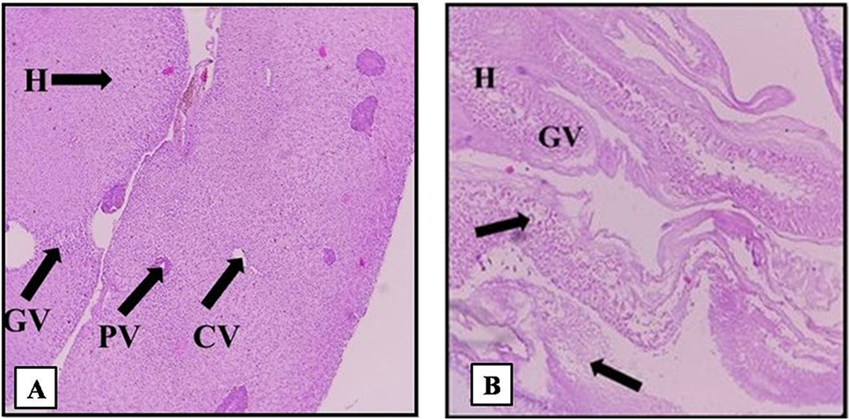
(A) Normal liver-normal architecture with no histological abnormalities was observed in liver of control fish. Normal hepatocytes can be seen (GV − Glycogen vacuole, H − Hepatocytes, CV − Central vein, PV − Portal vein). (B) Test liver − Complete structural disruption, severe necrosis of hepatocytes (arrow heads) with inflammatory cells were observed.
Current usage metrics show cumulative count of Article Views (full-text article views including HTML views, PDF and ePub downloads, according to the available data) and Abstracts Views on Vision4Press platform.
Data correspond to usage on the plateform after 2015. The current usage metrics is available 48-96 hours after online publication and is updated daily on week days.
Initial download of the metrics may take a while.


