Fig. 3
Download original image
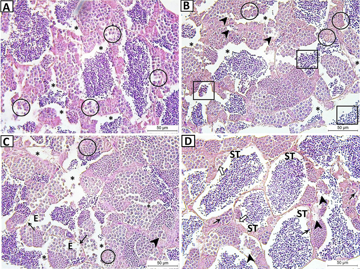
Testicular histopathology of 7.5 ppm mancozeb exposed samples. (A) Degenerative appereance of spermatogenic cells (encircled) in the disorganized tubules and increased intertubular spaces (asterisks). (B) Vacuole formations (arrowheads), degenerated spermatogenic cells (encirled), decreased sperm cells (squares) and increased separations (asterisks). (C) Edema (E), degenerated spermatogenic cells (encircled), hypertrophy of primary spermatocytes (arrowhead), thinned basement membrane (arrows) and separations (asterisks) (H&E staining). (D) Tubules without developing spermatogenic cells (ST), vacuolization (arrowheads), pyknotic nuclei (arrows) and karyolytic nuclei (white arrows). (H&E staining).
Current usage metrics show cumulative count of Article Views (full-text article views including HTML views, PDF and ePub downloads, according to the available data) and Abstracts Views on Vision4Press platform.
Data correspond to usage on the plateform after 2015. The current usage metrics is available 48-96 hours after online publication and is updated daily on week days.
Initial download of the metrics may take a while.